Figure 2
Partial SHOX duplications associated with various cases of congenital uterovaginal aplasia (MRKH syndrome): A tangible evidence but a puzzling mechanism
Daniel Guerrier* and Karine Morcel
Published: 24 March, 2021 | Volume 4 - Issue 1 | Pages: 001-008

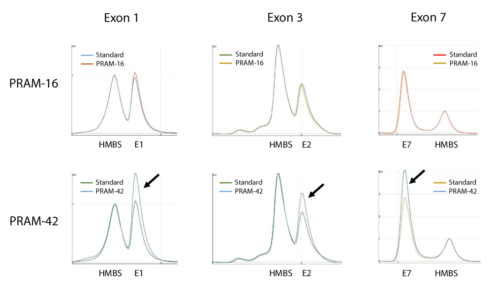
Figure 2:
DP/LC chromatograms for three different duplexes used in SHOX analysis. x-axis: retention time in min; y-axis: fluorescence intensity. Example of results obtained for quantification of copy number of SHOX exons 1, 3 and 7 in patients PRAM-16 and PRAM-42. In these experiments, a pool of 5 independent genomic DNAs was used as standard and the hydroxymethylbilane synthase (HMBS) gene, located at 11q23.2-qter, was used as an internal control. Profiles are superimposed and then normalized using the control amplicon for HMBS. Black arrows show the triple dosage of SHOX exons 1, 3 and 7 in patient PRAM-42. Primers sequences and location are summarized in Table 2.
Read Full Article HTML DOI: 10.29328/journal.jgmgt.1001006 Cite this Article Read Full Article PDF
More Images
Similar Articles
-
Partial SHOX duplications associated with various cases of congenital uterovaginal aplasia (MRKH syndrome): A tangible evidence but a puzzling mechanismDaniel Guerrier*,Karine Morcel. Partial SHOX duplications associated with various cases of congenital uterovaginal aplasia (MRKH syndrome): A tangible evidence but a puzzling mechanism. . 2021 doi: 10.29328/journal.jgmgt.1001006; 4: 001-008
Recently Viewed
-
Cytokine intoxication as a model of cell apoptosis and predict of schizophrenia - like affective disordersElena Viktorovna Drozdova*. Cytokine intoxication as a model of cell apoptosis and predict of schizophrenia - like affective disorders. Arch Asthma Allergy Immunol. 2021: doi: 10.29328/journal.aaai.1001028; 5: 038-040
-
Snow white: an allergic girl?Oreste Vittore Brenna*. Snow white: an allergic girl?. Arch Asthma Allergy Immunol. 2022: doi: 10.29328/journal.aaai.1001029; 6: 001-002
-
Impact of Latex Sensitization on Asthma and Rhinitis Progression: A Study at Abidjan-Cocody University Hospital - Côte d’Ivoire (Progression of Asthma and Rhinitis related to Latex Sensitization)Dasse Sery Romuald*, KL Siransy, N Koffi, RO Yeboah, EK Nguessan, HA Adou, VP Goran-Kouacou, AU Assi, JY Seri, S Moussa, D Oura, CL Memel, H Koya, E Atoukoula. Impact of Latex Sensitization on Asthma and Rhinitis Progression: A Study at Abidjan-Cocody University Hospital - Côte d’Ivoire (Progression of Asthma and Rhinitis related to Latex Sensitization). Arch Asthma Allergy Immunol. 2024: doi: 10.29328/journal.aaai.1001035; 8: 007-012
-
Gentian Violet Modulates Cytokines Levels in Mice Spleen toward an Anti-inflammatory ProfileSalam Jbeili, Mohamad Rima, Abdul Rahman Annous, Abdo Ibrahim Berro, Ziad Fajloun, Marc Karam*. Gentian Violet Modulates Cytokines Levels in Mice Spleen toward an Anti-inflammatory Profile. Arch Asthma Allergy Immunol. 2024: doi: 10.29328/journal.aaai.1001034; 8: 001-006
-
Effectiveness of levocetirizine in treating allergic rhinitis while retaining work efficiencyDabholkar Yogesh, Shah Tanush, Rathod Roheet, Paspulate Akhila, Veligandla Krishna Chaitanya, Rathod Rahul, Devesh Kumar Joshi*, Kotak Bhavesh. Effectiveness of levocetirizine in treating allergic rhinitis while retaining work efficiency. Arch Asthma Allergy Immunol. 2023: doi: 10.29328/journal.aaai.1001031; 7: 005-011
Most Viewed
-
Impact of Latex Sensitization on Asthma and Rhinitis Progression: A Study at Abidjan-Cocody University Hospital - Côte d’Ivoire (Progression of Asthma and Rhinitis related to Latex Sensitization)Dasse Sery Romuald*, KL Siransy, N Koffi, RO Yeboah, EK Nguessan, HA Adou, VP Goran-Kouacou, AU Assi, JY Seri, S Moussa, D Oura, CL Memel, H Koya, E Atoukoula. Impact of Latex Sensitization on Asthma and Rhinitis Progression: A Study at Abidjan-Cocody University Hospital - Côte d’Ivoire (Progression of Asthma and Rhinitis related to Latex Sensitization). Arch Asthma Allergy Immunol. 2024 doi: 10.29328/journal.aaai.1001035; 8: 007-012
-
Causal Link between Human Blood Metabolites and Asthma: An Investigation Using Mendelian RandomizationYong-Qing Zhu, Xiao-Yan Meng, Jing-Hua Yang*. Causal Link between Human Blood Metabolites and Asthma: An Investigation Using Mendelian Randomization. Arch Asthma Allergy Immunol. 2023 doi: 10.29328/journal.aaai.1001032; 7: 012-022
-
An algorithm to safely manage oral food challenge in an office-based setting for children with multiple food allergiesNathalie Cottel,Aïcha Dieme,Véronique Orcel,Yannick Chantran,Mélisande Bourgoin-Heck,Jocelyne Just. An algorithm to safely manage oral food challenge in an office-based setting for children with multiple food allergies. Arch Asthma Allergy Immunol. 2021 doi: 10.29328/journal.aaai.1001027; 5: 030-037
-
Snow white: an allergic girl?Oreste Vittore Brenna*. Snow white: an allergic girl?. Arch Asthma Allergy Immunol. 2022 doi: 10.29328/journal.aaai.1001029; 6: 001-002
-
Cytokine intoxication as a model of cell apoptosis and predict of schizophrenia - like affective disordersElena Viktorovna Drozdova*. Cytokine intoxication as a model of cell apoptosis and predict of schizophrenia - like affective disorders. Arch Asthma Allergy Immunol. 2021 doi: 10.29328/journal.aaai.1001028; 5: 038-040

If you are already a member of our network and need to keep track of any developments regarding a question you have already submitted, click "take me to my Query."


